
What you’ll find inside
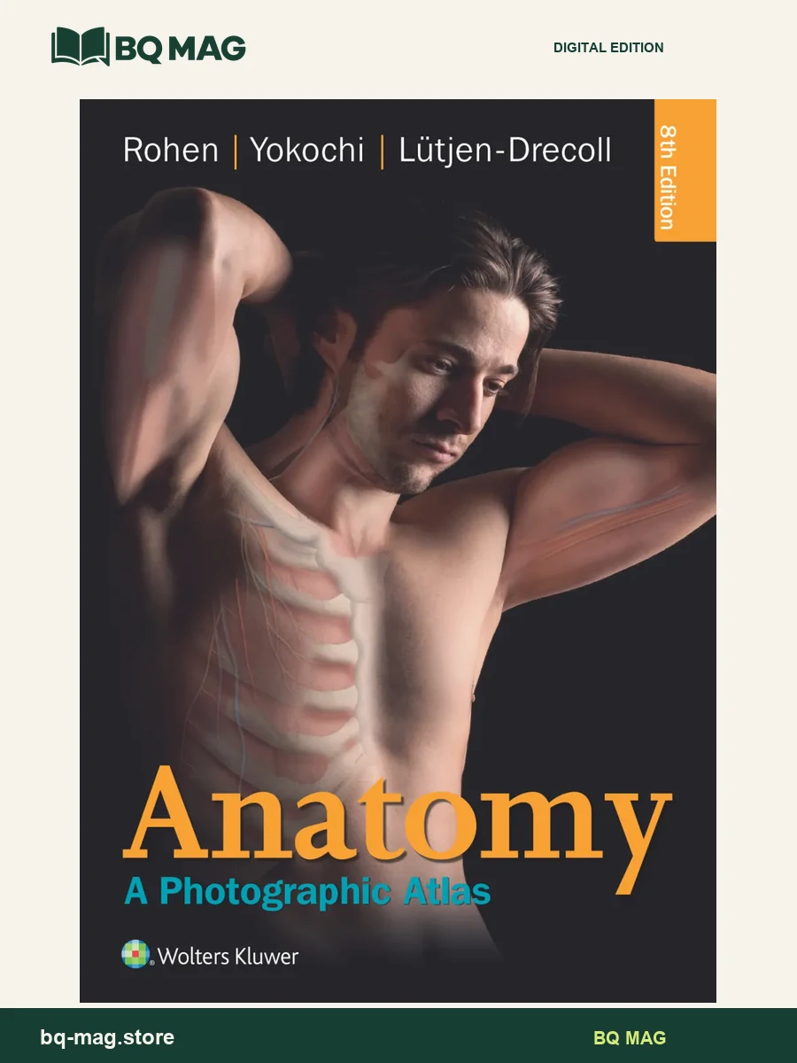
Anatomy: A Photographic Atlas, Eighth Edition, by Johannes W. Rohen, Chihiro Yokochi, and Elke Lütjen-Drecoll, is a visual reference organized around photographs of dissected human anatomy. Rather than relying only on simplified drawings, it places structures in their regional relationships and pairs photographic views with diagrams and imaging. That format can help anatomy students compare layers and landmarks as they prepare for laboratory work.
What makes this photographic anatomy atlas different?
The atlas presents anatomical structures through full-color cadaver dissections, supported by schematic drawings and diagnostic images. Its regional approach shows how tissues, vessels, nerves, organs, and neighboring structures occupy the same space. Readers can use the images to orient themselves before a practical session, then return to the relevant region when a structure is difficult to identify in a dissection or cross-sectional view.
How is the eighth edition arranged?
Material progresses from general anatomy to major body regions: head and neck, trunk, thoracic organs, abdominal organs, retroperitoneal organs, upper limb, and lower limb. Within these regions, the contents move among bones, joints, muscles, vessels, nerves, organs, and surface anatomy. This layout follows the way many courses teach regional anatomy and makes the atlas practical for reviewing one area at a time.
Who may benefit from this anatomy atlas?
The book is designed for readers who need a visual companion to anatomy study, including students in medicine and allied health fields. It can support preparation for dissection labs and help clarify spatial relationships that are hard to picture from text alone. Photographs are a study aid; learners should use them alongside current course materials, instructor guidance, and the lab specimens or models used in their program.
Edition, pages, and PDF preview
Bibliographic records identify the eighth edition and list 548 pages. The supplied product record is a PDF and its 562-page container count includes front matter. Check the eighth-edition designation against your reading list, since atlases with similar titles are available in several editions. Open the four-image preview tab to inspect sample pages and confirm that the file is the edition you need.
Browse more anatomy and physiology ebooks or return to the ebook catalog.
ISBN and edition details
ISBN for this edition (reference): 9781451193183
This identifier belongs to a published version of the matching title/edition. A format-specific ISBN for the supplied PDF has not been independently confirmed.


Victoria Persampiere –
An absolute must-have for medical school gross anatomy. Has real, labeled cadaver pictures from a variety of angles supplemented with diagrams of neurovasculature. If you’re only going to buy one ebook for anatomy, buy this one. (But I suggest you buy two: Rohens and Netters)
Marita –
Me parece mucho más fácil aprender anatomia con este tipo de imágenes, tb me sirve para usarlo de referencia cuando hago ecografias
NY –
questo è un vero atlante di anatomia con fotografie di cadaveri e visione straordinaria ed efficace di tutte le strutture anatomiche umane anche le vene e arterie più difficili da identificare però in italia è inutile… a medicina non sono previste dissezioni su cadavere quindi potete prenderlo so se vi piace la materia e se non vi accontentate degli atlanti pittorici tradizionali se volete capire chi siete e di “che pasta” siete fatti…è il vero ATLANTE!!!!
Ianna –
This ebook is amazing. Five million stars. It’s like having an anatomy cadaver lab in the comfort of your home minus the smells. So nice for studying in PA school. Worth every penny and then some. Of all the money I spent on Anatomy study resources this hands down was THE most important. Buy this ebook.
mrinal –
excellent ebook
Sal –
By far the best anatomy atlas for medical school, especially if you have lab practicals with the cadavers. Cadaver anatomy looks very different from the drawings, and a photographic atlas is a step in the right direction. Keep in mind though, it won’t substitute for the hours you put into studying the cadavers in person – no dissection or prosection you see in real life will ever be as clean and neat as Rohen’s photos.
Danny –
Highly recommended!!! Better than Netter for lab dissection. The cadavers photographs of anatomical structures really helps you learn the structure’s location on a cadaver. In terms of learning anatomical structure, this is the 2nd best resource (nothing bets learning anatomy from a cadaver). Prefect resource when you can’t or too lazy to go to Cadaver lab. The photographs instead of drawings, helps you get a better visualization of structures, and really helps for lab test. Each page of the atlas contains many labels and structures, so you don’t have to flip through page by page looking for a structure. Saves a lot of time!!! Very detailed photographs, photographs are the “cream of the crop” cadavers, illustrating the anatomical structure for easy visualization. So far I didn’t find any mistake or misspelling of any anatomical structures, however some names do vary from other atlas/text. Con: There absolutely no text or material content in this atlas, this is basically a true atlas listing out all the anatomical structures. No study guides or end of the chapter quiz (However comes with an online code that does come with online quizzes).